
Canine Ankylosis and Fixing CANTS - Dr. Maryam Hamid
Dr. Maryam Hamid
2 min read

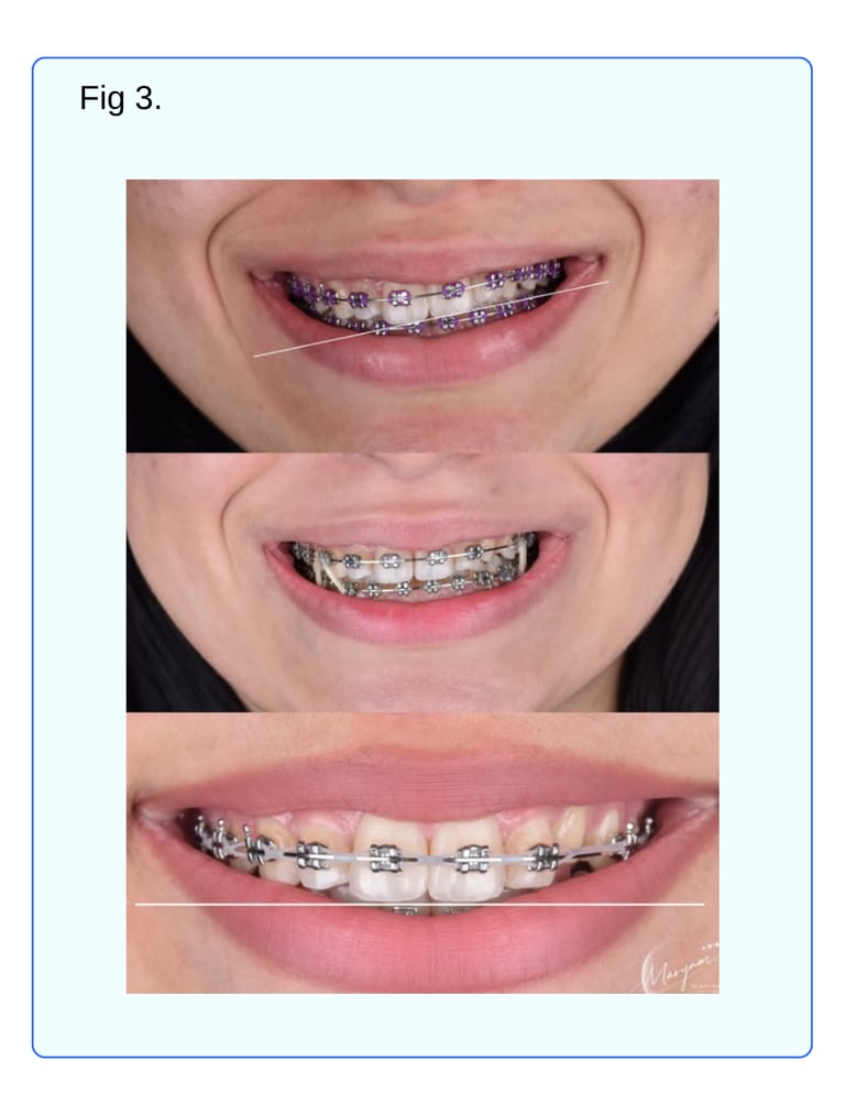
Chief Complaint (C.C.): A 17-year-old female patient presented with an unsatisfactory result from previous orthodontic treatment, complaining of minor lower crowding and a narrow smile.
Case Diagnosed During Orthodontic Treatment with “Progressive Replacement Ankylosis” and managed accordingly with appropriate Biomechanics.
Progressive Replacement Ankylosis is characterized by root resorption that leads to fusion of the tooth with surrounding bone, accompanied by simultaneous bone formation. This results in the gradual replacement of the tooth root by bone tissue.
Treatment steps :-
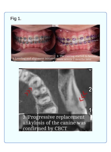
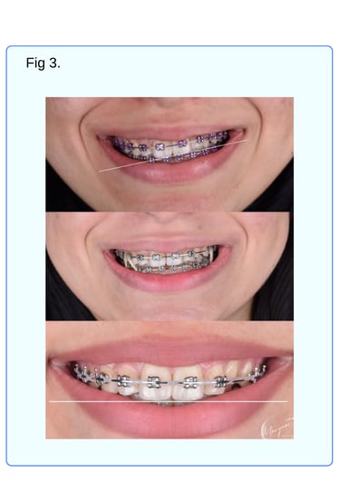
1. Leveling and Alignment
2. As canting became more apparent during treatment, the case was referred for CBCT imaging of UL3, UL4, and UL5 to identify which teeth were contributing to the canting and to confirm the presence of ankylosis.
3. After the diagnosis was confirmed, the canine was excluded from orthodontic treatment, as proceeding with the plan could lead to future fracture due to the thin root walls.
4. Leveling and alignment was restarted and continued until reaching heavy archwires (excluding UL3).
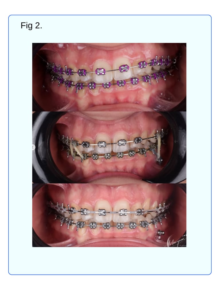
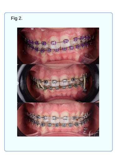
5. Extrusion of the upper left side was preferred over intrusion of the right side. Given the patient is a young female, a slightly visible gingival display is is acceptable and even favorable. A TAD was placed in the mandible between LL4 and LL5, applying a power chain from the TAD to the archwire (mesial and distal to LL4) to achieve intrusion of LL4/LL5 and simultaneous extrusion of the upper left segment using intermaxillary elastics (from TAD to UL4). Class I elastics were used on the right side.
6. After orthodontic treatment, the patient will be referred to a restorative dentist to manage the compromised UL3. The tooth will remain under observation. Should the resorption progress or ankylosis worsen, making the tooth structurally unsound, it will be replaced with a dental implant.
-Dr. Maryam Hamid Aldoori